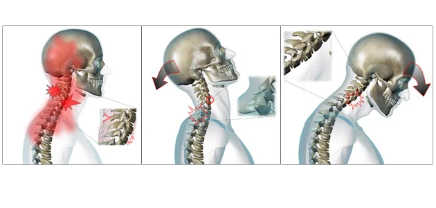
Chronic migraines are a debilitating condition that involves 15 or more migraines per month. That means a person is spending most of the time out of commission since migraines can often require a day or two to recover from.
The Effects of Cervical Spine Manipulation on Judo Athlete’s Grip Strength
Botelho, M.B., DC, Andrade B. B. MD, PhD. JMPT, November 2011

This article is straight forward and provides an excellent reference for those Upper Cervical chiropractors interested in athletics. The test population didn’t suffer from any particular condition, in fact they were male and female athletes from a nationally competitive judo team and cervical spinal manipulative therapy (or adjustments to the chiropractic community) made a statistically significant difference in their grip strength!
The study includes 18 athletes randomly assigned to either a treatment group or a sham adjustment group. The subjects where given 3 SMT within 3 weeks with a minimum of 36 hours between treatments. Grip strength was tested using a hydraulic dynamometer immediately before and after treatment. Grip strength improved in each hand pre and post intervention each time, the level of improvement was statistically significant, while no statistically significant difference was noted in the sham treatment group.
Alibhoy N. Resolution of Fibromyalgia Following Upper Cervical Chiropractic Care: A Case Study. J. Upper Cervical Chiropractic Research; June 20, 2011.

The case follows a 45 year old fibromyalgia patient with additional complaints of migraines, neck, upper back and low back pain, numbness in her fingers, bilateral sciatica, right knee pain, depression and duodenal ulcer. She had a history of two major cervical traumas and 11 car accidents, and had seen 5 different chiropractors in 12 years. Her activity levels were severely limited and she frequently used a wheel chair.
Knee Chest protocol was used and the patient was seen 79 times in 17 months with 47 adjustments performed to both atlas and axis.
At resolution of care the patient did not need the use of her wheel chair and self-reported fibromyalgia and left leg sciatica, right knee pain, chronic back pain and migraines had completely resolved. Right leg sciatica improved 98%. The patient was also no longer taking any of her previous routine medications.
Case studies are an invaluable starting point for more in-depth research.
The Effect of Upper Cervical or Sacroiliac Manipulation on Hip Flexion and Range of Motion
Pollard, DC, MS, Ward, PhD. JMPT 1998; 21(9);611-616

Your going to adjust my neck and it will help my hip feel better? How often have you heard this question in your office? The following study although a few years old points to a positive correlation and gives a compelling hypothesis for the connection.
This study compared the effectiveness of an upper cervical manipulation and a manipulation of the SI joint for increasing hip range of motion in 52 subjects ages 18-34. Testing methods where performed using a hand held digital electrogonimometer. The patients performed a straight leg raise before and after the treatment. The three treatment groups included just cervical manipulation, just SI joint manipulation (side posture) and the third received a sham adjustment of pressure on the mastoid process. Range of motion was tested prior to manipulation, the patient received one treatment and then range of motion was re-tested.
Both spinal manipulation groups demonstrated increased flexion of the hip however only the upper cervical manipulation increased hip flexion range of motion significantly.
The potential mechanism discussed is that of the tonic neck reflex. Changes in the muscle spindle output of the suboccipital muscles may cause reflexive proprioceptive changes to centers that control posture. The muscles of the pelvic girdle are some of our primary posture stabilizers.
Chronic neck pain, standing balance, and suboccipital muscle atrophy--a pilot study

McPartland JM, Brodeur RR, Hallgren RC, JMPT 1997 Jan;20(1):24-9
This study was completed at the University of Michigan and looked at 7 chronic neck pain patients and 7 controls.
The purpose of the study was to examine the relationship between chronic neck pain, standing balance and sub-occipital (the base of the skull) muscle atrophy.
Palpation was used to determine any somatic dysfunction (misalignment) of the upper cervical spine (top of the neck), a force platform was used to measure standing balance, and MRI was used to examine fatty infiltration (evidence of injury and disfunction) of the sub-occipital muscles.
The study found that chronic neck pain patients have almost twice the amount of somatic dysfunction (tenderness, asymmetry of joint position, restriction in range of motion, and tissue texture abnormality) as compared to normal subjects.
The greatest changes where noted at C0-C1 (where your skull meets your neck) joints and the authors concluded that this area needed the greatest amount of consideration during evaluation. (IE upper cervical chiropractic)
Further the study showed that chronic neck pain patients demonstrated a decrease standing balance using a force plate, and MR imaging indicated that they had increased atrophy of rectus capitus posterior minor and rectus capitus posterior major.
The authors also have a wonderful discussion with a compelling hypothesis of the far reaching implications of chronic neck pain…
“Somatic dysfunction can cause a sustained facilitation of motor neurons and reflex contraction of muscles, which may lead to impaired circulation and localized tissue ischemia, followed by atrophic changes in muscles and fatty degeneration. Muscle atrophy and degeneration have been associated with chronic pain. Muscles in the cervical region also contain a high density of muscle spindles… Atrophy of these muscles might reduce proprioceptive input into the dorsal horn of the spinal cord and higher centers… A reduction of proprioceptive input might result in facilitation of neural activity which is perceived by the patient as chronic pain.”
In review: Misalignment of the upper neck causes changes in the muscles and nerves in that area that affect your standing balance! Standing balance influences your posture, your posture contributes to breathing, hormone production, blood pressure, and more because it’s all connected! Further, the longer you have the neck pain the more negative changes develop.
Chronic Back Pain is Associated with Decreased Prefrontal and Thalamic Grey Matter Density
Apkarian V.A., et al. Journal of Neuroscience, Nov 2004, 24(46):10410-10415

This research was out of Northwestern University in Chicago Illinois in 2004. It was the first study to correlate chronic back pain (CBP) with decreased grey matter in the brain. As we work with patients every day, people who have chronic unremitting back pain for 1 year or more have an accelerated neurodegenerative process underway in their brain. If we are able to help them we are playing an active role in slowing that process!
The researchers studied 26 people with chronic back pain (unrelenting pain localized around lumbosacral area for greater than 1 year) and 26 control patients. They performed 2 different types of analysis for estimating global grey matter in the brain and adjusted statistics for age, gender, and type of pain (musculoskeletal and neurogenic/radicular).
Clinical Pearls:
Normal whole brain grey matter atrophy is 0.5% per year.
Atrophy caused by CBP was measured at 5-11% per year, the equivalent of 10-20 years of aging.
The reduction in grey matter was localized to the dorsolateral prefrontal cortex (DLPFC) and the thalamus. The DLPFC is responsible for inhibition of the orbitofrontal activity of the brain. The orbitofrontal area is responsible for perception of pain. The researchers then extrapolated that with loss of inhibition of the orbitofrontal areas of the brain, chronic pain suffers perceive increased pain.
Patients with neuropathic pain showed a greater loss of cortical grey matter.
Mechanisms of Musculoskeletal Pain
Bogduk N. The Journal of Orthopaedic Medicine 28(3) 2006
With three published texts and over 200 indexed articles , Nikolai Bogduk is one of the world’s foremost authorities on biomechanics of the spine and musculoskeletal pain, so when I came across this article I knew it would have some pertinent information that help us understand our patient’s pain.

Sample of Scalene Muscle trigger points and referred pain pattern
Key Points:
Pain transduction is ascribed to free or unencapsulated nerve endings with the following hierarchy of sensitivity; Periosteum, ligament, joint capsule, tendon, fascia, and muscle.
Reminder: that pain from a muscle is more commonly felt over the joint that that muscle moves.
How pain is created in the body: Mechanical or chemical stimuli affect free nerve endings in a peripheral nerve. Central transmission is then the term used for propagation of action potentials from the first order neurons (free nerve endings) to the second order neurons which form tracks in the spinal cord to higher centers in the brain and thalamus. Modulation then occurs in these tracks which involved intersegmental and descending pathways from the brainstem that inhibit and control the first synapse in this pain pathway. Physiologically it then follows that modulation is one of the mechanisms that upper cervical chiropractic helps control pain occurring almost anywhere in the body!
Sensory (afferent) nerves and Sympathetic nerves contribute to mechanisms of inflammation in the body. Chiropractic adjustments decrease sympathetic tone in the body, help to reduce inflammation and therefore pain.
Clinical Pearl: The next time you have an IME telling you that a patient has a ‘non-anatomical’ distribution of pain and therefore their pain is not genuine, you can also use this article to cite that ‘Ongoing pain sensitizes the central nervous system to produce larger areas of pain’ that may not follow classic anatomical distributions.

Not in the Portsmouth area? Click link above to find an Upper Cervical Chiropractor near you