Migraine or Headache? Knowing the Difference

Migraines are more than just bad headaches, as sufferers know all too well. So if you’ve been experiencing severe headaches, how can you know if this is actually a symptom of a migraine? Here are some more symptoms of migraines. Remember that people often exhibit different symptoms, so you may have some that are not listed here, but these are a few of the more common ones.

·         Sensitivity to light, sounds, and/or smells

·         Nausea and/or vomiting

·         Dizziness or vertigo

·         Visual disturbances (possibly including temporary loss of vision)

To learn more about the connection between head and neck injuries and migraines click the image below to request our complimentary e-book.

What Brings on a Migraine?

If your headaches turn out to be migraines, what can you do to prevent attacks, or keep them as far apart as possible? Here are a few practical suggestions:

·         Keep a diary – This will allow you to determine if certain foods, beverages, activities, or other triggers precede your attacks and avoid these.

·         Decrease stress levels – Migraines are often triggered by stress. You can reduce stress with a hot shower, a massage, some meditation, stretching, enjoying a hobby, or any other stress relief technique you can think of. Just remember to mark it in your journal if the stress relieving technique actually triggers a migraine.

·         See an upper cervical chiropractor

Why See an Upper Cervical Chiropractor?

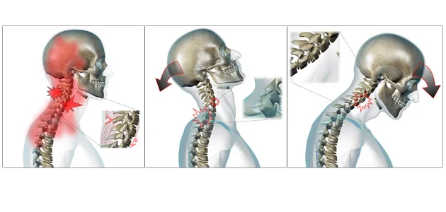
The fact is that your migraines may be the result of an upper neck misalignment. If you’ve ever been in an accident, suffered a sports injury, or even just hit your head, you may have a misalignment of the atlas (C1 vertebra). This isn’t something that would be corrected by a general chiropractor. It involves very specific measurements of the misalignment along with a very precise correction. At Arete Chiropractic, we use the Blair upper cervical technique to deliver just such an adjustment to our patients. In many case studies, upper cervical care has helped individuals suffering from migraines in as few as 1 to 2 adjustments. 

To schedule a consultation with Dr. Beebe or Dr. Evans click the button below or call 603-380-9184

Dizziness—What It Can Mean Following a Concussion

Dizziness can have many causes, but when the symptoms start after an injury to the head such as a concussion, the problem may actually be in your neck. What is the connection between head trauma and dizziness? Where can you look to for help in coping with dizziness after head or neck trauma?

Dizziness and concussions were connected again in the news lately as Cavs player Kevin Love was forced to leave a game after hitting his head on the court. He started to feel dizzy, was diagnosed with a concussion, and spent the rest of the game follow NBA protocols which involve leaving the court area to be in an environment that is quiet and distraction free.

That’s just one example of dizziness starting following an injury to the head. Many have experienced the same symptom after everything from a fall down the stairs to a car accident. If you feel your dizziness is connected to a recent or even a past head injury, what can you do?

To learn more about the connection between head and neck injuries and vertigo click the image below to request our complimentary e-book

Upper Cervical Care Provides Natural Benefits for Dizziness Sufferers

When a head injury is sustained, the neck is often jarred as well. If the upper vertebrae (the C1 and C2) misalign as a result of the injury, long lasting effects can occur, dizziness included. At Arete Chiropractic, we use the Blair method of upper cervical chiropractic to identify and correct this specific type of misalignment.

If you are worried about having your neck ‘popped’ following an injury, we can put your mind at ease. Upper cervical care is very different from general chiropractic. There is no ‘popping’ or ‘twisting’ involved, just a gentle correction that is long lasting. This allows any damaged caused by the misalignment to begin healing. If you are suffering from dizziness following a head or neck injury, come in for a consultation to find out what upper cervical chiropractic can do for you. 

To schedule a consultation with Dr. Beebe or Dr. Evans click the button below or call 603-380-9184

Headaches—Why the Neck Is a Good Place to Start Looking for Help

Headaches are known for the pain they cause to the head, but the genesis of many of these headaches could be in the neck. In fact, it was recently reported in an article on news-press.com (a subsidiary of USA Today) that up to 20% of headaches are cervicogenic—in other words, headaches caused by a disorder of the neck.

One of the things that cause pinched nerves and other issues in the neck that result in headaches is poor posture. In fact, whether it is being hunched over an iPad binge watching a TV show or sitting a desk at work with a monitor that isn’t positioned correctly, it’s surprising that more people are not facing this kind of headache.

To learn more about the connection between head and neck injuries and migraines download our complimentary e-book.

Help in Coping with Headache Pain

We want to make two recommendations for dealing with headaches. The first is to watch your posture at all times. Try sleeping on your back at night or on your side with a pillow between your knees rather than on your stomach. When sitting, make sure you are in a seat where your knees can bend at a 90-degree angle and your feet touch the floor (or use a footstool). Try to keep mobile devices at eye level by placing them on a stand on a table rather than leaning your head forward to look at them. Pay attention to your posture when standing and walking so that your head is not leaning forward. Proper posture will help reduce the occurrence of headaches.

Second, we encourage you to come in for a consultation at Arete Chiropractic. Even a slight misalignment of the upper neck can make proper posture feel unnatural. Plus, it can cause the rest of the spine to shift as the body compensates in order to keep your head straight. We use the Blair technique of upper cervical chiropractic to identify and correct such misalignments, and the result may be help for headache pain. 

To schedule a consultation with Dr. Beebe or Dr. Evans click the button below or call 603-380-9184

Imaging of the ligaments in the Upper Neck

Follow-Up MR Imaging of the Alar and Transverse Ligaments after Whiplash Injury:

A Prospective Controlled Study

Vetti N., Krakenes J. et al. American Journal of Neuroradiology 32: 1836-41, Nov 2011


Why do we do research? To answer questions?  Yet, how often do we seek out research that validates our paradigm?  My guess would be almost always.  Therein is the problem.  To be a true question, we must not think we already know the answer or outcome.  That was the reason that this study caught my eye.

This study was a prospective case controlled study that followed 91 symptomatic whiplash patients and 52 neck pain control patients for 1 year.  MR Imaging was done at baseline on neck pain patients and after the accident and on a 1 year follow up, results were also correlated with the neck disability index. 

The inclusion criteria for the whiplash included: MVA in the past 7 days with onset of neck pain within 48 hours.  No previous history of neck pain, and classified as WAD 1-2 (Whiplash Associated Disorders without any neurological signs, factures or dislocations).

The symptomatic control patients were included when they reported to an outpatient spine clinic with a history >3 months of neck pain with a non-traumatic onset.

The researchers concluded that the areas of high signal intensity (indicating inflammation/fibrosis/fat replacement) in the alar and transverse ligaments did not change significantly at baseline or at 1 year follow up for WAD patients.  Further the prevalence of the alar and transverse ligament high signal intensity did not differ significantly between traumatic and non-injured neck pain controls.

What the researchers concluded is that the alar and transverse ligament high signal intensity in patients with WAD1-2 observed in the first year after injury cannot be explained by the trauma.

Clinical Pearls:

“High signal intensity could theoretically be due to altered ligament function cause by neck pain.”

“Pain induced immobility causes morphologic changes in muscles, tendons and ligaments.”

“High signal intensity of the alar and transverse ligaments is also reported to be frequent in healthy non-injured persons without neck pain.”

Author’s Note: This study is a bit confounding however provides an interesting observation on neck pain related MR imaging. 

Grip Strength in Athletes improve with Cervical Adjusting

The Effects of Cervical Spine Manipulation on Judo Athlete’s Grip Strength

Botelho, M.B., DC, Andrade B. B. MD, PhD. JMPT, November 2011


This article is straight forward and provides an excellent reference for those Upper Cervical chiropractors interested in athletics.  The test population didn’t suffer from any particular condition, in fact they were male and female athletes from a nationally competitive judo team and cervical spinal manipulative therapy (or adjustments to the chiropractic community) made a statistically significant difference in their grip strength! 


The study includes 18 athletes randomly assigned to either a treatment group or a sham adjustment group.  The subjects where given 3 SMT within 3 weeks with a minimum of 36 hours between treatments.  Grip strength was tested using a hydraulic dynamometer immediately before and after treatment.   Grip strength improved in each hand pre and post intervention each time, the level of improvement was statistically significant, while no statistically significant difference was noted in the sham treatment group. 

Fibromyalgia responds to upper cervical care: A Case Study

Alibhoy N. Resolution of Fibromyalgia Following Upper Cervical Chiropractic Care: A Case Study. J. Upper Cervical Chiropractic Research; June 20, 2011.

The case follows a 45 year old fibromyalgia patient with additional complaints of migraines, neck, upper back and low back pain, numbness in her fingers, bilateral sciatica, right knee pain, depression and duodenal ulcer.   She had a history of two major cervical traumas and 11 car accidents, and had seen 5 different chiropractors in 12 years.  Her activity levels were severely limited and she frequently used a wheel chair. 

Knee Chest protocol was used and the patient was seen 79 times in 17 months with 47 adjustments performed to both atlas and axis. 

At resolution of care the patient did not need the use of her wheel chair and self-reported fibromyalgia and left leg sciatica, right knee pain, chronic back pain and migraines had completely resolved.  Right leg sciatica improved 98%.  The patient was also no longer taking any of her previous routine medications. 

 Case studies are an invaluable starting point for more in-depth research.    


Will how adjusting my neck affect my hip pain?

The Effect of Upper Cervical or Sacroiliac Manipulation on Hip Flexion and Range of Motion

Pollard, DC, MS, Ward, PhD.  JMPT 1998; 21(9);611-616

Your going to adjust my neck and it will help my hip feel better?   How often have you heard this question in your office?  The following study although a few years old points to a positive correlation and gives a compelling hypothesis for the connection.

This study compared the effectiveness of an upper cervical manipulation and a manipulation of the SI joint for increasing hip range of motion in 52 subjects ages 18-34.   Testing methods where performed using a hand held digital electrogonimometer.  The patients performed a straight leg raise before and after the treatment.  The three treatment groups included just cervical manipulation, just SI joint manipulation (side posture) and the third received a sham adjustment of pressure on the mastoid process.  Range of motion was tested prior to manipulation, the patient received one treatment and then range of motion was re-tested. 

Both spinal manipulation groups demonstrated increased flexion of the hip however only the upper cervical manipulation increased hip flexion range of motion significantly. 

The potential mechanism discussed is that of the tonic neck reflex.  Changes in the muscle spindle output of the suboccipital muscles may cause reflexive proprioceptive changes to centers that control posture.   The muscles of the pelvic girdle are some of our primary posture stabilizers.  

Is your neck pain affecting your balance?

Chronic neck pain, standing balance, and suboccipital muscle atrophy--a pilot study

 McPartland JM, Brodeur RR, Hallgren RC, JMPT 1997 Jan;20(1):24-9


This study was completed at the University of Michigan and looked at 7 chronic neck pain patients and 7 controls.

The purpose of the study was to examine the relationship between chronic neck pain, standing balance and sub-occipital (the base of the skull) muscle atrophy. 

Palpation was used to determine any somatic dysfunction (misalignment) of the upper cervical spine (top of the neck), a force platform was used to measure standing balance, and MRI was used to examine fatty infiltration (evidence of injury and disfunction) of the sub-occipital muscles.

The study found that chronic neck pain patients have almost twice the amount of somatic dysfunction (tenderness, asymmetry of joint position, restriction in range of motion, and tissue texture abnormality) as compared to normal subjects. 

The greatest changes where noted at C0-C1 (where your skull meets your neck) joints and the authors concluded that this area needed the greatest amount of consideration during evaluation.  (IE upper cervical chiropractic)

Further the study showed that chronic neck pain patients demonstrated a decrease standing balance using a force plate, and MR imaging indicated that they had increased atrophy of rectus capitus posterior minor and rectus capitus posterior major. 

The authors also have a wonderful discussion with a compelling hypothesis of the far reaching implications of chronic neck pain…

“Somatic dysfunction can cause a sustained facilitation of motor neurons and reflex contraction of muscles, which may lead to impaired circulation and localized tissue ischemia, followed by atrophic changes in muscles and fatty degeneration.  Muscle atrophy and degeneration have been associated with chronic pain.  Muscles in the cervical region also contain a high density of muscle spindles… Atrophy of these muscles might reduce proprioceptive input into the dorsal horn of the spinal cord and higher centers… A reduction of proprioceptive input might result in facilitation of neural activity which is perceived by the patient as chronic pain.”

In review:  Misalignment of the upper neck causes changes in the muscles and nerves in that area that affect your standing balance!  Standing balance influences your posture, your posture contributes to breathing, hormone production, blood pressure, and more because it’s all connected! Further, the longer you have the neck pain the more negative changes develop.  

Trigeminal neuralgia and migraine pain: One patient's response

Trigeminal neuralgia and migraine pain: One patient's response

Trigeminal Neuralgia and Chronic Migraine: One Patient's response to Blair Upper Cervical Care

Is your Back Pain shrinking your Brain?

Chronic Back Pain is Associated with Decreased Prefrontal and Thalamic Grey Matter Density

Apkarian V.A., et al.  Journal of Neuroscience, Nov 2004, 24(46):10410-10415

This research was out of Northwestern University in Chicago Illinois in 2004.  It was the first study to correlate chronic back pain (CBP) with decreased grey matter in the brain.  As we work with patients every day, people who have chronic unremitting back pain for 1 year or more have an accelerated neurodegenerative process underway in their brain.  If we are able to help them we are playing an active role in slowing that process!

The researchers studied 26 people with chronic back pain (unrelenting pain localized around lumbosacral area for greater than 1 year) and 26 control patients.  They performed 2 different types of analysis for estimating global grey matter in the brain and adjusted statistics for age, gender, and type of pain (musculoskeletal and neurogenic/radicular). 

Clinical Pearls:

Normal whole brain grey matter atrophy is 0.5% per year.

Atrophy caused by CBP was measured at 5-11% per year, the equivalent of 10-20 years of aging.

The reduction in grey matter was localized to the dorsolateral prefrontal cortex (DLPFC) and the thalamus.  The DLPFC is responsible for inhibition of the orbitofrontal activity of the brain.  The orbitofrontal area is responsible for perception of pain.  The researchers then extrapolated that with loss of inhibition of the orbitofrontal areas of the brain, chronic pain suffers perceive increased pain.

Patients with neuropathic pain showed a greater loss of cortical grey matter.

Is one leg shorter than the other?

Is one leg shorter than the other?

“approximately 90% of the population have anatomical difference in leg length, averaging 5.4mm(~3/16in)…whereas a difference of more than 20mm(~3/4in) is considered clinically significant in contributing to various musculoskeletal pathologies.”

Understanding Pain

Mechanisms of Musculoskeletal Pain

Bogduk N. The Journal of Orthopaedic Medicine 28(3) 2006

With three published texts and over 200 indexed articles , Nikolai Bogduk is one of the world’s foremost authorities on biomechanics of the spine and musculoskeletal pain, so when I came across this article I knew it would have some pertinent information that help us understand our patient’s pain.

Sample of Scalene Muscle trigger points and referred pain pattern

Sample of Scalene Muscle trigger points and referred pain pattern

Key Points:

Pain transduction is ascribed to free or unencapsulated nerve endings with the following hierarchy of sensitivity; Periosteum, ligament, joint capsule, tendon, fascia, and muscle.

Reminder: that pain from a muscle is more commonly felt over the joint that that muscle moves.

How pain is created in the body: Mechanical or chemical stimuli affect free nerve endings in a peripheral nerve.  Central transmission is then the term used for propagation of action potentials from the first order neurons (free nerve endings) to the second order neurons which form tracks in the spinal cord to higher centers in the brain and thalamus.  Modulation then occurs in these tracks which involved intersegmental and descending pathways from the brainstem that inhibit and control the first synapse in this pain pathway.  Physiologically it then follows that modulation is one of the mechanisms that upper cervical chiropractic helps control pain occurring almost anywhere in the body!

Sensory (afferent) nerves and Sympathetic nerves contribute to mechanisms of inflammation in the body.  Chiropractic adjustments decrease sympathetic tone in the body, help to reduce inflammation and therefore pain.

Clinical Pearl: The next time you have an IME telling you that a patient has a ‘non-anatomical’ distribution of pain and therefore their pain is not genuine,  you can also use this article to cite that ‘Ongoing pain sensitizes the central nervous system to produce larger areas of pain’ that may not follow classic anatomical distributions.